Normal Fetal Outcome in a Case of Prenatally Diagnosed Placental Mesenchymal Dysplasia
* Gharib M;
Bahadorian S;
-
* Gharib M: Department of Pathology, Faculty of Medicine, Mashhad University of Medical Sciences, Mashhad, Iran
-
Bahadorian S: Department of Pathology, Faculty of Medicine, Mashhad University of Medical Sciences, Mashhad, Iran
-
Sep 25, 2026 |
-
Volume: 3 |
-
Issue: 1 |
-
Views: 33 |
-
Downloads: 44 |
Abstract
Background: Placental Mesenchymal Dysplasia (PMD) is a rare placental anomaly characterized by abnormal mesenchymal tissue proliferation and is associated with increased risks of fetal complications and intrauterine death.
Case Presentation: We report the case of a 23-year-old gravida 3, para 1 + 1 (one live birth, one stillbirth due to meconium aspiration) woman who was prenatally suspected of having PMD or a molar pregnancy based on a routine ultrasound at 23 weeks’ gestation. Her prenatal care included serial ultrasound examinations to monitor fetal growth and well-being.
Outcomes and Follow-up: Spontaneous labor occurred at 37 weeks’ gestation. A healthy male infant weighing 3,120 g with Apgar scores of 9 at 1 minute and 10 at 5 minutes was delivered by cesarean section. Postpartum pathological examination of the placenta confirmed the diagnosis of PMD.
Introduction
Placental mesenchymal dysplasia (PMD) is a rare placental anomaly first described by Moscoso et al. [1,2] as stem-villus hyperplasia. It is characterized by placentomegaly and sonographic features that can mimic a molar pregnancy, such as “grapelike” vesicles. However, PMD has distinct pathological findings: unlike molar pregnancies, it lacks trophoblastic proliferation and instead shows aneurysmal dilatation of fetal surface vessels and dilated stem villi.
In many cases, PMD is diagnosed incidentally during routine prenatal ultrasound examinations, as in the present case. The etiology of PMD remains unclear, but a mesodermal origin is suspected. The defining pathological feature is mesenchymal hyperplasia within stem villi [3]. PMD has also been reported in association with other conditions characterized by mesodermal proliferation. Enlarged stem villi in PMD often contain acid mucopolysaccharides, which are typically present in normal chorionic mesoderm. Moreover, approximately 23% of PMD cases co-occur with Beckwith–Wiedemann syndrome (BWS), suggesting a possible etiological link.
Because PMD can closely resemble a molar pregnancy on prenatal ultrasound (due to placentomegaly and cystic, vesicular areas), accurate differentiation is critical: the clinical course and prognosis differ markedly. Unlike molar pregnancies, PMD may be compatible with a healthy fetal outcome.
Case Presentation
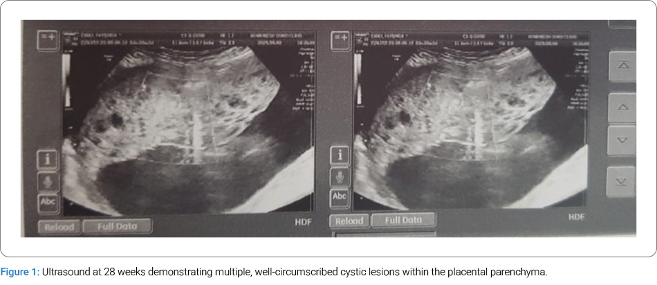
A 23-year-old gravida 3, para 1 + 1 (one live birth, one stillbirth) woman presented to our antenatal clinic for a routine ultrasound at 19 weeks’ gestation. The initial ultrasonographic findings suggested a molar pregnancy. Further investigations were performed, including serial sonographic evaluations at 23 and 28 weeks’ gestation, and an amniocentesis at 20 weeks, which showed a normal karyotype.
These assessments revealed a progressively enlarged placenta with multiple well-circumscribed cystic areas throughout the parenchyma, consistent with placental mesenchymal dysplasia (Figure 1).
Course and Outcome
At 37 weeks’ gestation, the patient underwent a cesarean section, delivering a healthy male infant weighing 3,120 g, with Apgar scores of 9 at 1 minute and 10 at 5 minutes.
Macroscopic Placental Pathology
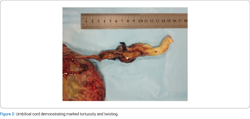
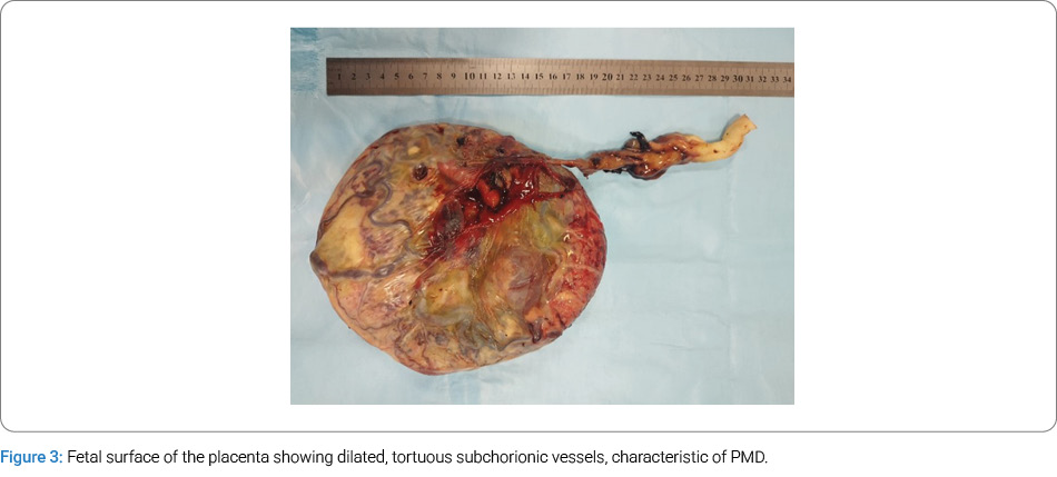
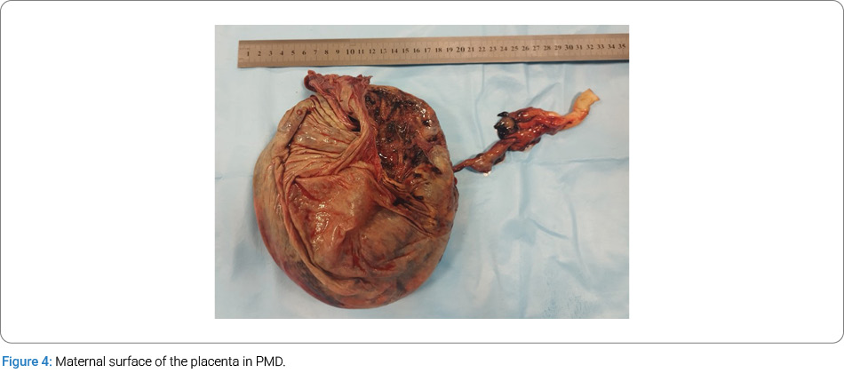
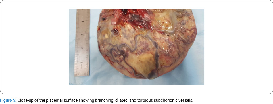
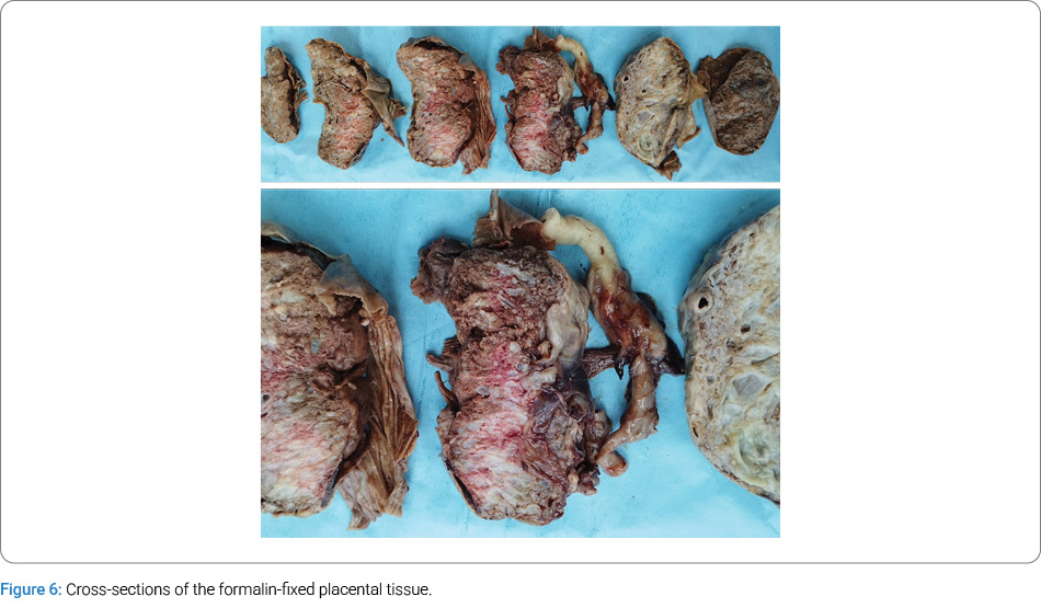
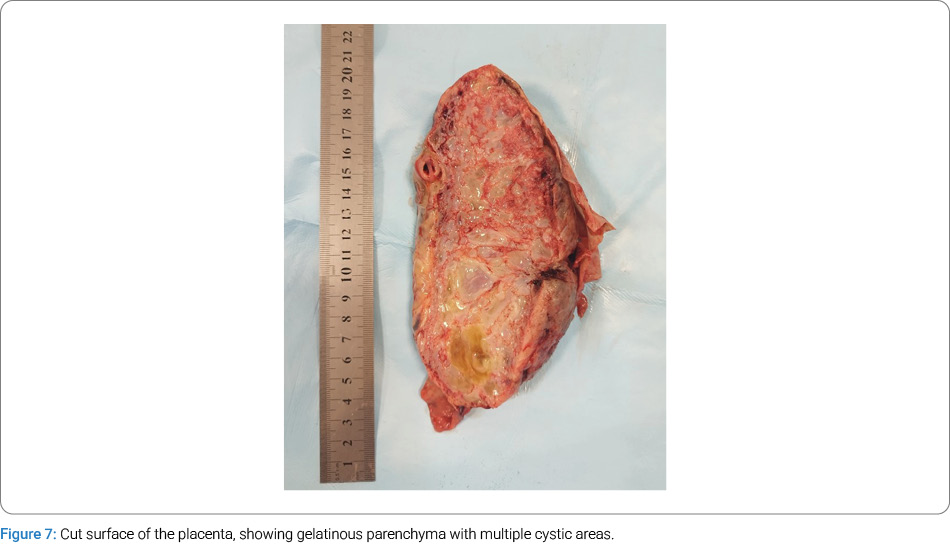
The placenta was markedly enlarged, weighing 875 g and measuring 20 cm × 18 cm × 4 cm. The umbilical cord was 18 cm long and 1 cm–2.3 cm thick, with three vessels, eccentric insertion, and marked tortuosity (Figure 2). The fetal surface exhibited dilated and tortuous subchorionic vessels (Figure 3). On sectioning, the parenchyma appeared gelatinous and contained multiple cystic areas (Figure 4–Figure 7).
Microscopic Findings
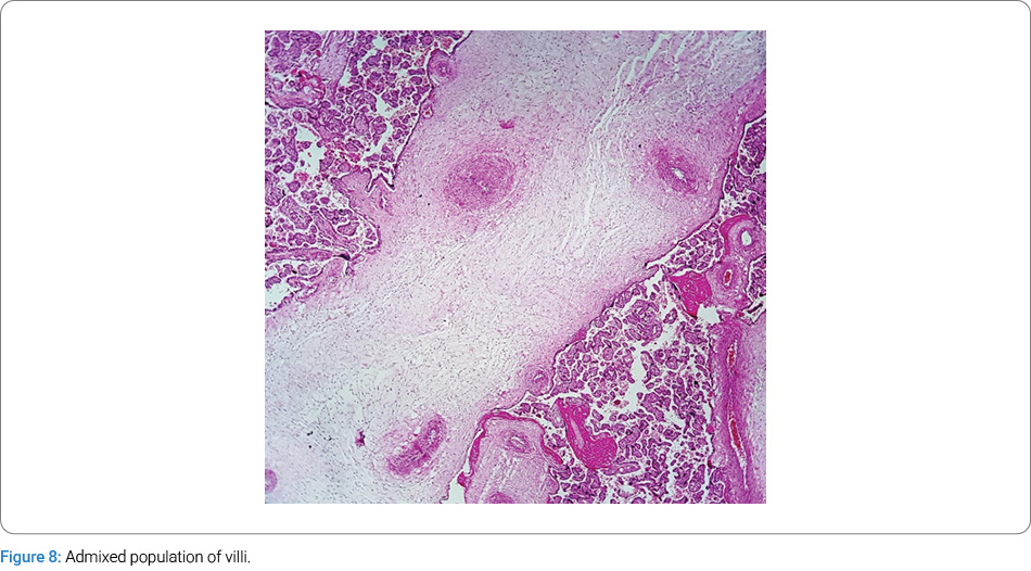
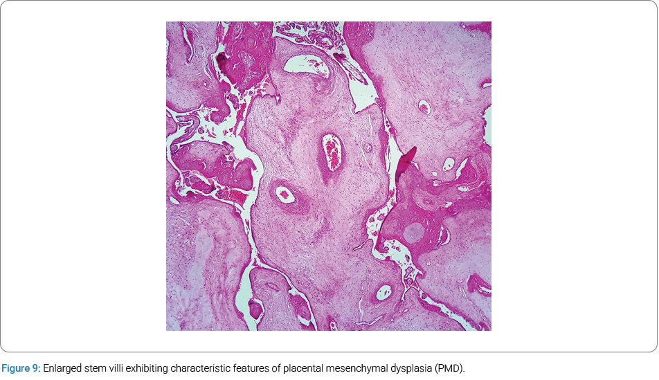
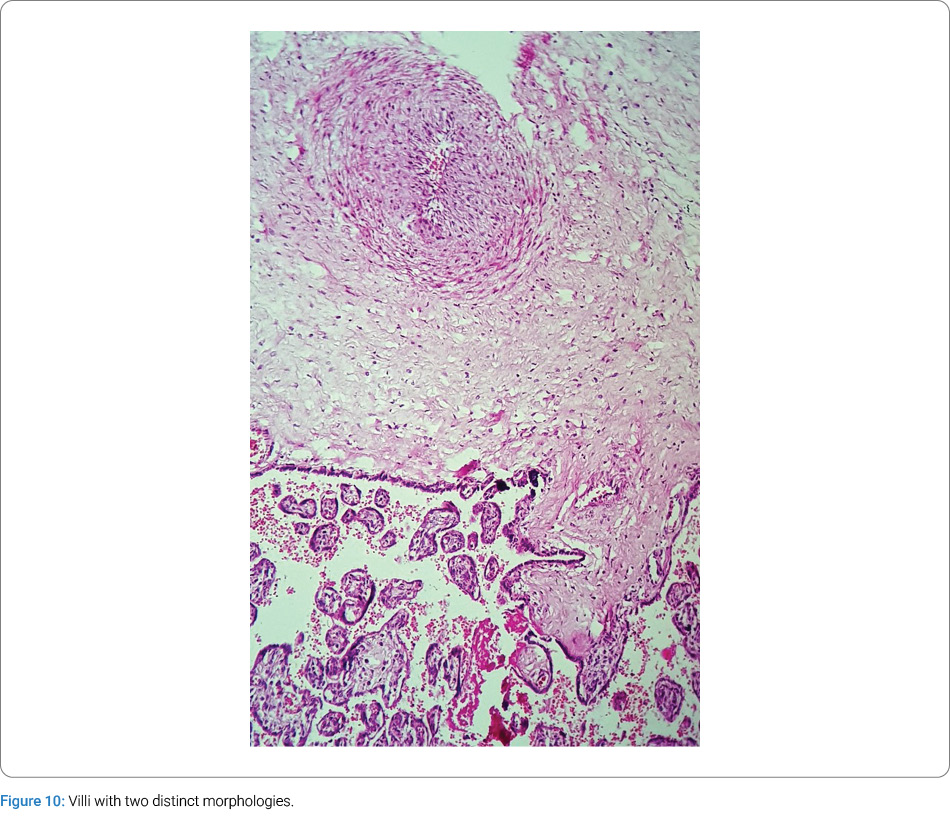
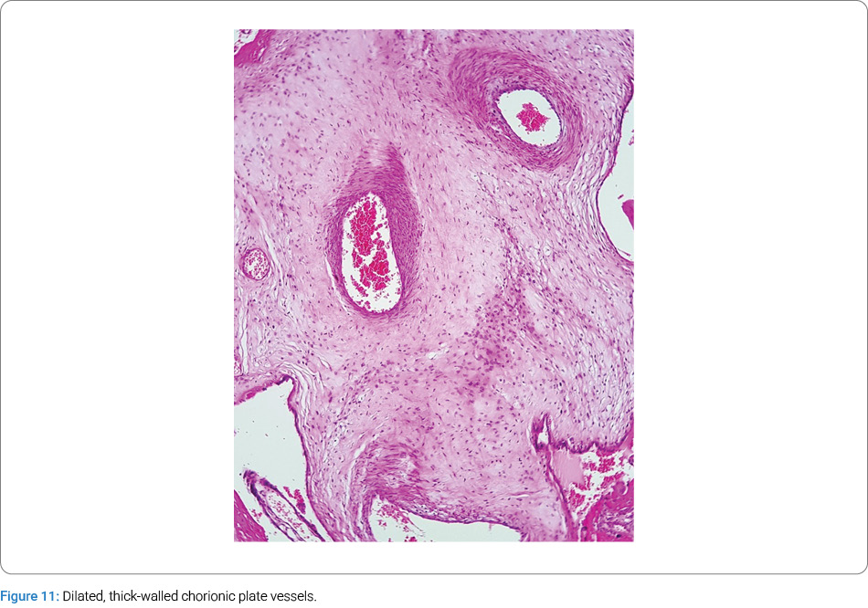
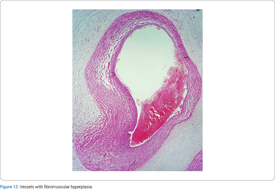
Histological examination revealed two distinct populations of villi. The first appeared largely unremarkable and appropriate for gestational age (Figure 8). The second consisted of markedly enlarged stem villi displaying features characteristic of PMD, including hydropic stroma, central cistern formation, thick-walled vessels, and myxoid stroma (Figure 9–Figure 10). Additional sections (Figure 11–Figure 12) demonstrated dilated, thick-walled chorionic plate vessels with fibromuscular hyperplasia. There was a conspicuous absence of trophoblastic hyperplasia. Furthermore, intermediate villi exhibited changes reminiscent of chorangiomatosis, with abnormal vascular branching and dilation.
Discussion
PMD is likely underdiagnosed and underreported, which makes its true incidence uncertain. Current estimates suggest a prevalence of approximately 0.02%. PMD is frequently associated with Beckwith–Wiedemann Syndrome (BWS), a disorder characterized by somatic overgrowth and a predisposition to embryonal tumors. The BWS locus on chromosome 11p15.5 involves key genes, such as those encoding Insulin-like Growth Factor-2 (IGF-2) [4]. Research suggests that altered regulation of IGF-2 (for example, loss of maternal suppression) may contribute to BWS and possibly to PMD. Indeed, about 23% of PMD cases are reported in conjunction with BWS, supporting the hypothesis of a shared etiology.
Epigenetic dysregulation of imprinted genes on 11p15.5 — including CDKN1C (p57^KIP2), H19, IGF-2, and KVLQT — has been implicated in PMD [5]. Many of our insights into the molecular basis of PMD derive from research on BWS, suggesting that defects in cell-cycle regulation may drive the abnormal mesenchymal proliferation observed in PMD [6,7]. Accurate diagnosis is essential for appropriate pregnancy management. Differentiating PMD from other placental pathologies, such as Partial Hydatidiform Mole (PHM) and BWS, is critical. Imaging modalities, including ultrasound and MRI, play an indispensable role in establishing the diagnosis and excluding other potential conditions [8–10].
Conclusion
Placental mesenchymal dysplasia is a rare but benign placental disorder, characterized by cystic changes within the placental parenchyma. Although its etiology and long-term fetal outcomes remain incompletely understood, precise diagnosis is crucial to distinguish it from more ominous pathologies. When a cystic placenta is observed on prenatal ultrasound, PMD should be considered in the differential diagnosis. Close fetal monitoring is essential to detect potential complications and to avoid unnecessary pregnancy termination. Routine prenatal ultrasound, supplemented by advanced imaging techniques such as MRI when indicated, can facilitate the identification and assessment of PMD. Further longitudinal studies are needed to delineate its natural history and prognosis.
Acknowledgment
This case report was presented as a poster at the 27th Iranian Annual Congress of Pathology and Laboratory Medicine.
Conflict of Interest
The authors declare no potential conflicts of interest with respect to the research, authorship, and/or publication of this article. Informed consent was obtained for this publication.
References
- Taga S, Haraga J, Sawada M, Nagai A, Yamamoto D, Hayase R. A case of placental mesenchymal dysplasia. Case Rep Obstet Gynecol. 2013;2013:265159.
- Moscoso G, Jauniaux E, Hustin J. Placental vascular anomaly with diffuse mesenchymal stem villous hyperplasia. A new clinico-pathological entity? Pathol Res Pract. 1991;187(2– 3):324–328.
- Parveen Z, Tongson-Ignacio JE, Fraser CR, Killeen JL, Thompson KS. Placental mesenchymal dysplasia. Arch Pathol Lab Med. 2007;131(1):131–137.
- Weksberg R, Shen D, Fei YL, Song QL, Squire J. Disruption of insulin–like growth factor 2 imprinting in Beckwith– Wiedemann syndrome. Nat Genet. 1993;5(2):143–150.
- Arizawa M, Nakayama M. Suspected involvement of the X chromosome in placental mesenchymal dysplasia. Congenit Anom (Kyoto). 2002;42(4):309–317.
- Weksberg R, Shuman C, Beckwith JB. Beckwith–Wiedemann syndrome. Eur J Hum Genet. 2010;18(1):8–14.
- Cohen MC, Roper EC, Sebire NJ, Stanek J, Anumba DOC. Placental mesenchymal dysplasia associated with fetal aneuploidy. Prenat Diagn. 2005;25(3):187–192.
- Paradinas FJ, Sebire NJ, Fisher RA, Rees HC, Foskett M, Seckl MJ, et al. Pseudo-partial moles: placental stem vessel hydrops and the association with Beckwith-Wiedemann syndrome and complete moles. Histopathology. 2001;39(5):447–454.
- Maher ER, Reik W. Beckwith–Wiedemann syndrome: imprinting in clusters revisited. J Clin Invest. 2000;105(3):247– 845.
- Ping AJ, Reeve AE, Law DJ, Young MR, Boehnke M, Feinberg AP. Genetic linkage of Beckwith–Wiedemann syndrome to 11p15. Am J Hum Genet. 1989;44(5):720–723.
Cite this article
Bahadorian S, Gharib M. Normal Fetal Outcome in a Case of Prenatally Diagnosed Placental Mesenchymal Dysplasia. Global J Gynecol Obstet Res. 2026;3(1):1–5.
Keywords
Placental mesenchymal dysplasia; Fetal outcome; Prenatal diagnosis; Hydatidiform mole
Copyright
© 2026 Gharib M. This is an open access article distributed under the terms of the Creative Commons Attribution 4.0 International License (CC BY-4.0).