Pediatric Streptococcal Sepsis Complicated by Primary Psoas Abscess and Cardiac Involvement: A Case Report
* Huda Raja;
Midhat Farooq Malik;
Aleena;
Anza Muhammad Mohsin;
-
* Huda Raja: Department of Pediatrics, Dow University of Health Sciences, New Labour Colony Nanakwara, Karachi, Pakistan
-
Midhat Farooq Malik: Department of Pediatrics, Dow University of Health Sciences, New Labour Colony Nanakwara, Karachi, Pakistan
-
Aleena: Department of Pediatrics, Dow University of Health Sciences, New Labour Colony Nanakwara, Karachi, Pakistan
-
Anza Muhammad Mohsin: Department of Pediatrics, Dow University of Health Sciences, New Labour Colony Nanakwara, Karachi, Pakistan
-
Oct 20, 2026 |
-
Volume: 3 |
-
Issue: 1 |
-
Views: 5 |
-
Downloads: 2 |
Abstract
Background: Primary psoas abscess is a rare but potentially serious condition in children, often resulting from hematogenous spread of infection. In tuberculosis-endemic countries like Pakistan, Mycobacterium tuberculosis is frequently considered the primary culprit. However, pyogenic causes must not be overlooked. This case report describes an unusual presentation of psoas abscess secondary to streptococcal sepsis, further complicated by cardiac dysfunction, underscoring the importance of early diagnosis and multidisciplinary care.
Case Presentation: A 10-year-old previously healthy boy presented with a one-month history of fever, right scapular swelling, and pain radiating from the right hip to the leg. Imaging studies confirmed abscesses in the scapular and psoas regions. Investigations ruled out tuberculosis and other common etiologies. Persistently elevated Anti-Streptolysin O (ASO) titers indicated streptococcal sepsis. The patient was managed with intravenous antibiotics, heart failure therapy, and long-term secondary prophylaxis with benzyl penicillin. After six weeks of treatment, he achieved complete clinical and functional recovery.
Conclusion: To our knowledge, this is the first reported pediatric case in Pakistan involving a streptococcal psoas abscess with cardiac complications. This case highlights the necessity of including pyogenic infections in the differential diagnosis of pediatric hip pain and demonstrates the effectiveness of timely diagnosis and collaborative treatment strategies.
Abbreviations
ASO: Anti-Streptolysin O; MRI: Magnetic Resonance Imaging; TB: tuberculosis; CT: Computed Tomography
Introduction
Psoas abscess, although uncommon, should be considered in children presenting with nonspecific symptoms such as fever, abdominal pain, or difficulty in walking. The condition arises from bacterial infection of the iliopsoas compartment and may be classified as either primary or secondary. Primary psoas abscesses typically occur without an identifiable adjacent infection and are thought to arise from hematogenous spread from a distant site [1]. Secondary abscesses are more common in adults and usually result from direct extension of nearby infections such as osteomyelitis, appendicitis, or gastrointestinal tract disease.
Staphylococcus aureus is the most commonly isolated organism in primary cases, accounting for up to 88% of infections [2,3]. Other pathogens, including Streptococcus and Escherichia coli, have been implicated less frequently, accounting for 4.9% and 2.8% of primary cases, respectively [3]. In tuberculosis-endemic regions, Mycobacterium tuberculosis remains a frequent cause of iliopsoas abscesses, especially in children [1].
The psoas muscle’s anatomical proximity to various structures and its rich blood supply predisposes it to infectious seeding. In this case, the patient developed a primary psoas abscess secondary to streptococcal bacteremia and subsequently developed cardiac complications. This case report adds to the limited literature on pediatric pyogenic psoas abscess and highlights the diagnostic complexity and management of such cases.
Case Presentation
A 10-year-old boy presented to the emergency department with a history of intermittent fever for the past month, accompanied by swelling over the right scapular area and progressive pain radiating from the right hip to the leg. He had no prior history of trauma, sore throat, skin infections, or recent travel. There was no known exposure to tuberculosis or previous significant illnesses.
On physical examination, the child was febrile (39.2°C) with a mildly elevated heart rate of 110 beats per minute. Other vital signs were stable. A 6-inch soft, fluctuant, non-tender swelling was noted over the right scapular region. The child had difficulty bearing weight on his right leg, with pain exacerbated by hip extension. Tenderness was elicited over the right hip and inguinal area, and the range of motion was significantly restricted.
Initial laboratory investigations revealed leukocytosis (WBC: 19,500/μL with neutrophilic predominance), elevated CRP (112 mg/L), and a markedly raised ESR (110 mm/hr). Blood cultures showed no growth, and HIV screening was negative. Given the region’s endemic status, tuberculosis (TB) was ruled out using GeneXpert testing and gastric aspirate analysis.
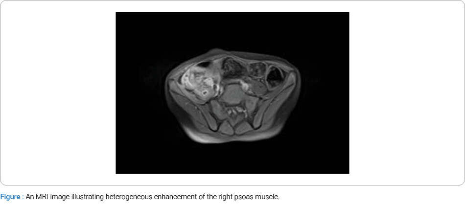
Ultrasound of the scapular swelling revealed a loculated fluid collection with internal septations (3.9 × 2.1 × 3.1 cm, approximately 13.7 mL), suggestive of an abscess. Additionally, abdominal ultrasound showed a collection in the right psoas muscle (8.4 × 3.6 × 4.1 cm, 66 mL), consistent with a psoas abscess. There were no signs of joint effusion in the right hip or knee. An MRI of the lumbosacral spine confirmed the presence of myositis and a right-sided psoas abscess, with no involvement of spinal vertebrae or discs. MRI also revealed thickening of the right psoas and iliacus muscles, with heterogeneous hyperintense signal on T2-weighted and STIR images and isointense signal on T1-weighted images, extending from L3 to the lesser trochanter. The right psoas muscle measured 24 × 6 mm at the level of L5-S1 (Figure).
The patient began to experience shortness of breath and developed bilateral pedal edema during hospitalization. A transthoracic echocardiogram revealed left ventricular systolic dysfunction with an ejection fraction of 45%, prompting initiation of anti-failure treatment per cardiology recommendations.
Cultures from aspirated fluid remained sterile, likely due to prior antibiotic exposure. However, three consecutive ASO titers were significantly elevated (> 1440 IU/mL), strongly supporting the diagnosis of streptococcal sepsis. Management was conservative, coordinated among pediatric medicine, infectious diseases, cardiology, and pediatric surgery. The patient received intravenous ceftriaxone and vancomycin for four weeks. As a preventive measure against rheumatic heart disease and recurrent streptococcal infections, intramuscular benzyl penicillin prophylaxis (monthly) was initiated, planned for one year.
Clinical improvement was noted within two weeks: the fever subsided, and the child began walking with minimal support. By the end of the fourth week, he regained normal mobility. Repeat ultrasound after six weeks demonstrated a significant reduction in psoas abscess size—from 66 mL to 6 mL. Follow-up echocardiography at three months showed complete resolution of ventricular dysfunction, leading to discontinuation of anti-failure medications.
Discussion
Psoas abscesses were once primarily linked to spinal tuberculosis, but modern studies have broadened the microbial spectrum and highlighted the growing prevalence of pyogenic cases, especially in immunocompetent pediatric patients [4,5]. Primary psoas abscess is rare in children and often misdiagnosed due to its nonspecific symptoms. The classic triad of fever, limp, and back pain, described as early as 1881, is seen in fewer than one-third of cases [6]. Instead, children might present with more subtle symptoms such as malaise, hip pain, or difficulty walking, which can easily be mistaken for conditions like septic arthritis, osteomyelitis, or appendicitis [7].
Diagnosing psoas abscess in regions where tuberculosis is endemic can be particularly challenging, as TB is often the presumed cause. However, once TB was excluded, we turned to other potential pyogenic organisms, with the elevated ASO titers ultimately guiding us to the diagnosis of streptococcal sepsis, a rare but well-documented source of psoas abscesses [3].
This case was further complicated by the development of heart failure symptoms during hospitalization, which illustrates the potential for systemic manifestations in streptococcal infections. This highlights the critical need for cardiovascular assessments in patients with systemic streptococcal infection. Despite sterile cultures, possibly due to prior antibiotic therapy, elevated ASO titers suggested streptococcal bacteremia, a rare but significant cause of deep tissue infections, such as psoas abscesses [8]. This case underscores the importance of considering such uncommon complications.
Imaging techniques, particularly MRI, were instrumental in confirming the diagnosis for us. In some studies, MRI was compared with CT, and it was concluded that MRI provides superior soft-tissue resolution and allows for more precise detection of abscess walls and adjacent tissue involvement, especially when evaluating intra-abdominal abscesses [9]. As CT was unavailable in this case due to limited resources at a government hospital, we were unable to reach a conclusion. This should be evaluated further to review false-negative cases.
The trend toward conservative treatment, especially for moderate-sized abscesses in hemodynamically stable patients, has been well documented in case series, such as those by van den Berge and Shields et al. [10,11]. Non-surgical management, particularly with antibiotic monotherapy, has been found to yield favorable results, especially in cases stemming from skeletal iliopsoas abscesses rather than gastrointestinal or urinary sources [12]. Our patient’s clinical improvement with antibiotics alone further supports this approach.
Conclusion
This case highlights a rare instance of streptococcal sepsis leading to primary psoas abscess with associated cardiac dysfunction in a pediatric patient. In regions where tuberculosis is prevalent, pyogenic etiologies must not be overlooked, especially when initial tests are negative. Timely imaging, guided antimicrobial therapy, and multidisciplinary care were key to the positive outcome. The case adds valuable insight to the limited pediatric literature on non-tuberculous psoas abscesses and supports conservative management in suitable cases.
Conflict of Interest
The authors declare no potential conflicts of interest with respect to the research, authorship, and/or publication of this article. Informed consent was obtained for this publication.
References
- Christin L, Sarosi GA. Pyomyositis in North America: case reports and review. Clin Infect Dis. 1992;15(4):668–677.
- Mallick IH, Thoufeeq MH, Rajendran TP. Iliopsoas abscesses. Postgrad Med J. 2004;80(946):459–462.
- Ricci MA, Rose FB, Meyer KK. Pyogenic psoas abscess: worldwide variations in etiology. World J Surg. 1986;10(5):834–843.
- Idris I, Aburas M, Ibarra Martinez F, Osei-Kuffuor E, Adams K, Dizadare T, et al. Primary Psoas Abscess in a Pediatric Patient: A Case Report. Cureus. 2022;14(6):e26206.
- Taiwo B. Psoas abscess: a primer for the internist. South Med J. 2001;94(1):2–5.
- Karlı A, Belet N, Danacı M, Avcu G, Paksu Ş, Köken Ö, et al. Iliopsoas abscess in children: report on five patients with a literature review. Turk J Pediatr. 2014;56(1):69–74.
- Qureshi NH, O’Brien DP, Allcutt DA. Psoas abscess secondary to discitis: a case report of conservative management. J Spinal Disord. 2000;13(1):73–76.
- Gordin F, Stamler C, Mills J. Pyogenic psoas abscesses: noninvasive diagnostic techniques and review of the literature. Rev Infect Dis. 1983;5(6):1003–1011.
- Shields D, Robinson P, Crowley TP. Iliopsoas abscess--a review and update on the literature. Int J Surg. 2012;10(9):466–469.
- van den Berge M, de Marie S, Kuipers T, Jansz AR, Bravenboer B. Psoas abscess: report of a series and review of the literature. Neth J Med. 2005;63(10):413–416.
- Yacoub WN, Sohn HJ, Chan S, Petrosyan M, Vermaire HM, Kelso RL, et al. Psoas abscess rarely requires surgical intervention. Am J Surg. 2008;196(2):223–227.
- López VN, Ramos JM, Meseguer V, Pérez Arellano JL, Serrano R, Ordóñez MAG, et al.; GTI-SEMI Group. Microbiology and outcome of iliopsoas abscess in 124 patients. Medicine (Baltimore). 2009;88(2):120–130.
Cite this article
Malik MF, Aleena, Mohsin AM, Raja H. Pediatric Streptococcal Sepsis Complicated by Primary Psoas Abscess and Cardiac Involvement: A Case Report. Global J Pediat. 2026;3(1):1–3.
Keywords
Streptococcal psoas abscess; Streptococcal sepsis; Cardiac dysfunction
Copyright
© 2026 Huda Raja. This is an open access article distributed under the terms of the Creative Commons Attribution 4.0 International License (CC BY-4.0).